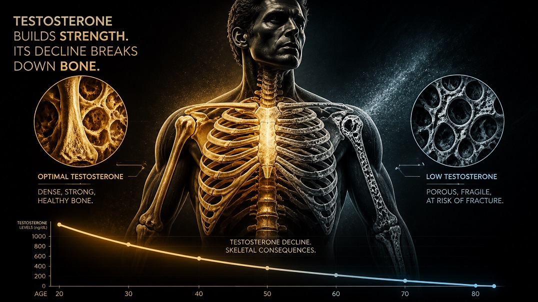
Bone loss is not a condition most men think about until it becomes a problem — and by the time it becomes a problem, it often already has for years. Osteoporosis is widely understood as a women’s health issue, and the assumption that men are largely exempt from significant bone loss has been consequential: men are diagnosed later, treated less aggressively, and suffer worse outcomes following osteoporotic fractures than women with comparable bone density loss.
Testosterone is one of the primary regulators of bone density in men. It’s not the only one — estrogen, which is produced from testosterone by aromatization, plays an equally important role — but the hormonal architecture of male skeletal health is fundamentally dependent on sex hormone status. Men with low testosterone lose bone at accelerated rates, and the fracture consequences of that bone loss are serious. A hip fracture in a man over 70 carries a one-year mortality rate approaching 30 percent — a statistic that puts the skeletal consequences of hormonal decline in a very different frame than the conversation usually occupies.
This article covers how testosterone and estrogen maintain bone density in men, what happens to bone when testosterone declines, what the risk looks like quantitatively, and what interventions — hormonal, nutritional, and exercise-based — have the strongest evidence for protecting bone health in men as they age.
How Sex Hormones Regulate Bone in Men
Bone is living tissue in constant turnover. Osteoclasts break down old bone; osteoblasts build new bone. In a healthy hormonal environment, this remodeling cycle is roughly in balance, maintaining bone mineral density and structural integrity. Sex hormones — both testosterone and estrogen — are critical regulators of this balance, acting through receptors expressed on both osteoclasts and osteoblasts.
Testosterone’s Direct Role in Bone Maintenance
Androgen receptors are expressed on osteoblasts — the bone-building cells — and testosterone directly stimulates osteoblast activity and survival. It supports the production of growth factors involved in bone matrix formation, including IGF-1, and promotes the differentiation of mesenchymal stem cells toward osteoblasts rather than adipocytes (fat cells). Testosterone also suppresses osteoclast activity, reducing the rate of bone resorption.
Studies on men with hypogonadism — whether from primary testicular failure, pituitary disorders, or iatrogenic testosterone suppression (as in androgen deprivation therapy for prostate cancer) — consistently show accelerated bone loss and higher fracture rates. Men on androgen deprivation therapy, who experience profound testosterone suppression, can lose five to ten percent of bone mineral density in the first year alone — a loss that takes decades to accumulate under normal age-related decline. This dramatic natural experiment in testosterone deprivation provides some of the clearest evidence for testosterone’s role in male bone health.
The Surprising Role of Estrogen in Male Bone
Estrogen — produced in men through aromatization of testosterone in fat tissue, muscle, and bone itself — turns out to be at least as important as testosterone for male bone density, and possibly more so for certain aspects of bone maintenance. This finding, which emerged from studies of men with estrogen receptor mutations or aromatase deficiency, was initially surprising but is now well established.
Research has found that in older men, estradiol levels are more strongly correlated with bone mineral density and fracture risk than testosterone levels, and that the bone loss associated with declining testosterone is partly mediated through the resulting decline in estrogen production. This has a practical implication: men with very low body fat — who have less peripheral aromatization — may show more significant bone effects from testosterone decline than men who carry more body fat, despite the latter group’s testosterone often being lower due to aromatase-driven conversion.
It also means that overly aggressive estrogen suppression in men on TRT — through aromatase inhibitors used to manage elevated estradiol — carries a genuine bone risk if it drives estrogen too low. The balance between testosterone and estrogen matters for skeletal health, not testosterone alone.
The Timeline of Male Bone Loss
Men reach peak bone mass in their late 20s to early 30s. From that point, bone density is relatively stable through the 30s and begins declining gradually from the late 30s onward. The rate of loss accelerates with age — particularly after 65 — and is significantly influenced by testosterone status throughout.
Men lose bone more slowly than women during midlife, partly because they don’t experience the abrupt estrogen withdrawal of menopause. But they live longer than the traditional narrative has acknowledged, and by the 70s and 80s, male bone loss has accumulated to a degree that produces clinically significant fracture risk. Estimates suggest that one in four men over 50 will experience an osteoporotic fracture in their lifetime — a figure that remains largely unknown to most men and underappreciated by many clinicians.
The relationship between testosterone decline and bone loss isn’t a future risk for men in their 50s — it’s a process that is already underway. The decade from 45 to 55, during which testosterone begins its most consequential decline for many men, is also a period when bone maintenance depends heavily on sustaining adequate sex hormone levels. Decisions made about hormonal health, exercise, calcium and vitamin D intake, and other bone-relevant variables during this window have long consequences.
Risk Factors That Compound Testosterone-Related Bone Loss
Low testosterone accelerates bone loss, but it rarely operates in isolation. Several other factors common in the population of men most relevant to this site compound the skeletal risk.
Vitamin D Deficiency
Vitamin D is essential for calcium absorption from the gut and for the bone mineralization process through which osteoblasts produce mature, dense bone. Deficiency is extremely common — estimates suggest that 40 to 70 percent of men in northern latitudes are deficient or insufficient — and it directly compounds the bone effects of low testosterone by impairing the mineralization that makes new bone structurally sound. The testosterone-bone relationship operates most effectively in an adequate vitamin D environment. Our article on how sunlight and vitamin D affect testosterone covers the vitamin D picture and how to address it.
Physical Inactivity and Sedentary Behavior
Bone responds to mechanical loading — the compressive and tensile forces generated by weight-bearing activity and muscle contraction. Without adequate mechanical stimulation, bone remodeling shifts toward net loss even in men with adequate sex hormone levels. Sedentary men show lower bone density than active men at comparable ages and hormonal status, and immobilization — whether from injury or extended bed rest — produces rapid and dramatic bone loss that illustrates how dependent skeletal maintenance is on mechanical loading. This means that the sedentary lifestyle associated with desk-based work compounds both the testosterone and bone-related risks simultaneously.
Alcohol and Smoking
Both alcohol and smoking are independent risk factors for bone loss in men. Alcohol impairs osteoblast function, reduces calcium absorption, and suppresses testosterone — compounding the hormonal and direct skeletal effects simultaneously. Smoking reduces estrogen levels (through accelerated estrogen metabolism), impairs blood supply to bone, and is consistently associated with lower bone mineral density and higher fracture rates. Men who drink heavily and smoke are carrying a significant additive bone risk on top of any testosterone-related acceleration of loss.
Certain Medications
Several commonly prescribed medication classes accelerate bone loss and deserve mention for men in the relevant age range. Glucocorticoids (corticosteroids like prednisone) are the most potent drug-related bone loss risk, capable of producing significant density reductions within months of use. Proton pump inhibitors (PPIs) — extremely widely used for acid reflux — reduce calcium absorption and are associated with modest increases in fracture risk with long-term use. Some antidepressants, anticonvulsants, and androgen deprivation therapies also carry bone risks. Men on these medications should discuss bone health monitoring with their physician.
Assessing Bone Density: What Testing Looks Like
Bone mineral density is measured by DEXA scan (dual-energy X-ray absorptiometry) — a low-radiation imaging study that takes approximately 10 to 20 minutes and provides density measurements at the hip and lumbar spine, the two most clinically important sites for fracture risk. Results are reported as T-scores: the number of standard deviations above or below the mean for a young adult reference population. A T-score above -1 is normal; between -1 and -2.5 is osteopenia (below-normal density, increased fracture risk); below -2.5 is osteoporosis.
Current guidelines recommend DEXA screening for men over 70 and for younger men with significant risk factors — including confirmed hypogonadism, glucocorticoid use, prior low-trauma fracture, or other clinical risk factors. In practice, many men with low testosterone in their 40s and 50s who have had multiple years of suboptimal hormone levels would benefit from earlier screening, though this isn’t universally recommended. Discussing DEXA with a physician alongside testosterone evaluation is a reasonable step for men with confirmed low T.
The FRAX tool — a validated fracture risk calculator developed by the World Health Organization — combines bone density with clinical risk factors to estimate 10-year fracture probability and is widely used to guide treatment decisions. It’s available online and provides a useful context for understanding individual risk beyond the T-score alone.
Protecting Bone Health: What the Evidence Supports
The interventions with the strongest evidence for protecting male bone health address the hormonal, mechanical, and nutritional dimensions of bone maintenance simultaneously.
Resistance Training and Impact Exercise
Weight-bearing exercise — particularly resistance training with progressive loading and high-impact activities like running and jumping — is the most potent non-pharmacological intervention for bone density preservation and improvement. The mechanical loading of resistance training directly stimulates osteoblast activity through the piezoelectric effect in bone tissue, producing new bone formation in response to compressive forces. Studies on resistance training and bone density in middle-aged and older men consistently find meaningful preservation or modest improvement in density at the hip and spine — the sites of highest fracture consequence.
The exercises with the strongest bone stimulus are those involving heavy axial loading — squats and deadlifts load the spine and hip directly and produce the largest osteogenic stimulus of any common resistance exercises. Our article on the best exercises for boosting testosterone naturally covers the training approach that supports both hormonal and skeletal health simultaneously.
Calcium and Vitamin D
Adequate calcium intake — approximately 1,000 to 1,200 mg daily for men over 50, from food sources primarily — provides the mineral substrate for bone formation. Dairy products, leafy greens, fortified foods, and calcium-rich fish like sardines are the primary dietary sources. Vitamin D, as discussed above, is essential for calcium absorption and bone mineralization; most men at northern latitudes require supplementation to maintain adequate levels, particularly through winter months. The combination of adequate calcium and vitamin D is the nutritional foundation on which both exercise and hormonal interventions act.
Testosterone Replacement and Bone
For men with confirmed hypogonadism, testosterone replacement therapy has demonstrated meaningful improvements in bone mineral density, particularly at the lumbar spine. Studies on TRT and bone typically show density increases of three to ten percent over two to three years — improvements that translate into meaningful reductions in fracture risk. The effect is most pronounced in men with the lowest starting testosterone and most significant baseline bone loss, and appears to plateau as bone density normalizes over several years of treatment.
Our articles on the benefits of TRT and monitoring your health on TRT cover the bone density dimension of testosterone treatment in more detail. For men considering TRT partly on the basis of bone protection, DEXA monitoring before and during treatment is a reasonable way to track the skeletal response.
Questions Men Ask About Testosterone and Bone Density
How much bone density can low testosterone cause me to lose?
The rate of bone loss associated with low testosterone varies by severity and duration of deficiency. Men on androgen deprivation therapy — who experience severe, medically induced testosterone suppression — can lose five to ten percent of bone density in the first year, illustrating the extreme end of the spectrum. Men with moderate age-related testosterone decline lose bone more gradually, but over a decade the accumulated loss can move a man from normal density to osteopenia, meaningfully increasing fracture risk. The impact of testosterone on bone is cumulative and silent until fracture occurs, which is why monitoring bone health alongside testosterone is clinically appropriate.
At what age should men start thinking about bone density?
Earlier than most men do. Peak bone mass is achieved in the late 20s, and the factors that influence it — exercise habits, calcium and vitamin D intake, sex hormone status, smoking and alcohol — matter throughout the 30s and 40s even though bone loss hasn’t yet become clinically apparent. Men with confirmed low testosterone in their 40s should discuss bone density assessment with their physician. For most men without specific risk factors, formal screening begins later — but the lifestyle habits that protect bone are worth establishing long before a DEXA scan is indicated.
Can you rebuild lost bone, or only prevent further loss?
Both are possible, though rebuilding bone is slower and more limited than preventing loss. Testosterone treatment in hypogonadal men, resistance training, and adequate calcium and vitamin D can all produce modest increases in bone mineral density — particularly in men with significant prior loss and at the lumbar spine, which is more responsive to intervention than the hip. Pharmacological interventions for osteoporosis — bisphosphonates, denosumab, and others — can produce more substantial density improvements but are typically reserved for men with established osteoporosis or high fracture risk. The practical emphasis for most men is preventing the loss that produces these more aggressive treatment scenarios rather than reversing it after the fact.
Does dairy consumption actually matter for bone health?
The evidence on dairy and fracture risk is more nuanced than the longstanding public health messaging suggests. Calcium intake, of which dairy is a primary source in Western diets, is genuinely important for bone mineral density. But the relationship between dairy consumption specifically and fracture outcomes in epidemiological studies is less consistent than expected, and countries with high dairy consumption don’t show uniformly lower fracture rates than those with lower consumption. The current evidence supports adequate calcium intake — primarily from food sources, with dairy being an efficient option for those who tolerate it — as part of a broader approach to bone health that includes exercise, vitamin D, and hormonal optimization. Dairy alone is not a bone health strategy.
Is osteoporosis reversible once it develops?
Significant improvements in bone density are possible in men with established osteoporosis, particularly with appropriate pharmacological treatment alongside hormonal optimization and exercise. Bisphosphonates and other agents can produce density increases of five to ten percent over two to three years and meaningfully reduce fracture risk even in men with significant baseline loss. However, the structural changes in bone architecture that accompany advanced osteoporosis — changes in trabecular microstructure that aren’t fully captured by density measurements — are less reversible than density loss alone. Earlier intervention produces better outcomes than waiting for significant architectural deterioration.
Should I get a DEXA scan if my doctor hasn’t recommended one?
If you have confirmed low testosterone, are over 50, have multiple risk factors for bone loss (smoking, significant alcohol use, glucocorticoid treatment, sedentary lifestyle, low body weight), or have experienced a low-trauma fracture, discussing DEXA with your physician is a reasonable and well-supported request. Current guidelines underscreen men relative to the actual burden of osteoporotic fractures in the male population, and men who advocate for earlier assessment are acting on sound evidence. A DEXA scan is low-risk, relatively inexpensive, and provides information that directly influences the management of both bone health and the hormonal picture.